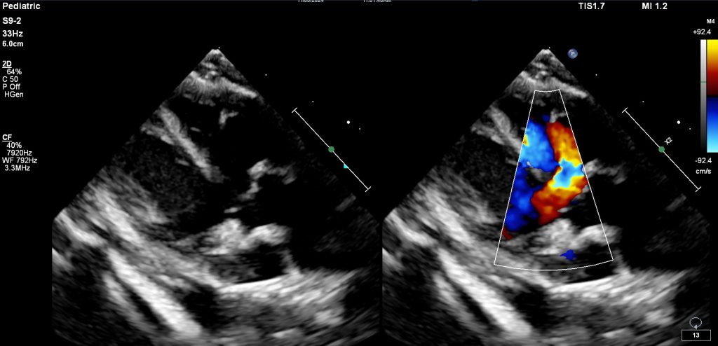
Sweet Etta James was only 6 days old when she was diagnosed. Her vet heard a heart murmur and her owner noticed occasional labored breathing. An echocardiogram revealed the hole in her heart. She was prescribed benazepril (an ACE inhibitor) to reduce fluid build up, and she will be monitored closely. Fortunately, she has no clue! She was happily dancing around the clinic and giving us kisses.


The ventricles are the muscular pumping chambers of the heart. The right ventricle pumps blood to the lungs where it picks up oxygen and returns to the left side of the heart, and the left ventricle pumps the oxygenated blood to the body. Normally, the left and right ventricles are separated by a muscular septum. If the septum is incompletely formed during development in utero, this results in a connection between the right and left chambers. This hole is called a ventricular septal defect, or VSD. The higher pressure on the left side of the heart drives blood across the hole into the right heart, causing it to recirculate through the lungs.

Blood moving through the hole generates a heart murmur that can be heard when listening with a stethoscope. Over time, the extra blood flow through the lungs can cause high blood pressure (pulmonary hypertension) or fluid build up in the lungs (congestive heart failure), resulting in trouble breathing. Medications can be prescribed to relieve the symptoms. There are some surgical treatment options, but these should be given careful consideration on an individual basis.
We use technologies like cookies to store and/or access device information. Consenting to these technologies will allow us to process data such as browsing behavior or unique IDs on this site. Not consenting or withdrawing consent, may affect some features and functions of this site.
